It is estimated that 10% of the population have diabetes, which is a condition where the body does not handle glucose in the blood appropriately, resulting in periods of prolonged hyperglycaemia (increased blood sugar levels). This affects the small blood vessels in the body and causes them to leak or become narrower, thereby compromising blood supply to the end organs that they supply.
The retina of our eyes is equivalent to the film in a pre-digital era camera (the eye is often thought of as being similar to a camera). It is also one of the vital organs that are affected by diabetes, resulting in diabetic retinopathy. It is estimated that 80% of diabetics will have diabetic retinopathy after 10 years of being diagnosed. Of course, many diabetics have the condition way before the actual diagnosis is made.
Diabetic retinopathy can be briefly divided into 2 broad categories namely, non-proliferative or background retinopathy and the more advanced stage of proliferative diabetic retinopathy, where new vessels grow as a result of insufficient oxygen supply to the retina.
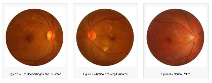
The challenge is to screen for the disease so that retinopathy can be detected at the non-proliferative stage. A retinal examination or a retinal photograph will show up small leaks from the blood vessels in the form of dot or blot like bleeding areas in the retinal layer (see Figure 1), or leakage of fatty substances from the vessels which will be seen as yellowish deposits (see Figure 2).
 If fluid leaks into the retinal layer in sufficient quantities as to cause a swelling of the retina, this is called macula edema, and this can be detected only when examined by an eye specialist because it requires a 3-dimensional perception of the retina.
Patients should be aware that diabetic retinopathy may not cause symptoms but they are at risk of visual loss. It is very worthwhile to screen for the condition because prompt treatment can prevent loss of vision and prevent blindness in up to 90% of cases. Of course, patients with diabetes should control their levels of blood sugar, blood pressure and cholesterol; but when afflicted with diabetic retinopathy and require treatment to prevent vision loss, they should know that the treatment is highly effective.
Treatment consists of laser placed onto the retina. The procedure is done under topical (eye drop) anaesthesia as a day case and patients can go home on the same day. The treatment may need to be repeated in certain cases and the patients must be properly counseled in order that they do not default on the treatment.
If detected early enough, laser treatment alone will suffice to prevent visual loss. Additional treatment modalities for diabetic retinopathy include injections of certain chemicals (that retard the formation of new blood vessels) into the eye, or surgery within the eye (vitrectomy).
Patients should be screened at least once a year, even when they do not have any visual symptoms, and be aware that in doing so, they are also being checked for the other eye complications that a diabetic is at increased risk of, such as earlier development of cataract and higher risk of developing glaucoma.
